Molecular Probes Cell Imaging Guide with Protocols
This mobile app is designed to help you find fluorescent dyes, reagents and protocols for cell biology related fluorescence microscopy applications.
Molecular Probes mobile app is great for:
• Selecting science protocols for labeling organelles and cellular structures such as: Golgi, mitochondria, nucleus, endoplasmic reticulum, plasma membrane, actin, tubulin, peroxisomes, autophagasomes, endosomes and lysosomes without Spectraviewer.
• Finding Alexa Fluor® labeled mouse, goat and rabbit secondary antibodies for immunocytochemistry and immunohistochemistry applications on Zeiss, Leica, Olympus and Nikon microscope systems.
• Choosing calcium ion indicators, cell health assays, fluorophores and pH dyes for neuroscience, neurobiology and cancer research.
• Discovering which Invitrogen, Gibco or Ambion cell health, cell culture, or reactive dye kits are best for benchtop cytometry, tissue staining, or biotin streptavidin conjugation for microscopic labeling and detection.
• Remote access to troubleshoot issues of: photobleaching, background fluorescence, and autofluorescence using signal enhancers, signal amplification, phalloidin conjugates, and click chemistry.
Product selections are available in the following categories:
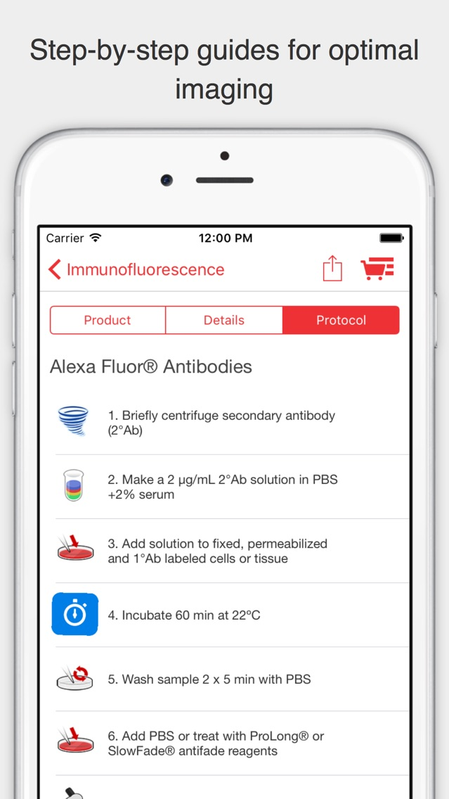
Immunofluorescence
Cell Proliferation
DNA Stains
GFP & RFP
Trafficking
Apoptosis
Oxidative Stress
Cell Viability
Organelle Stains
Cytoskeleton
Antifades
Antibody Labeling
Cell Cycle
Autophagy
Ion Indicators
Protein Labeling
Epitope Tags
RNA Biology
Protein Labeling
Essentials
Stem Cell Biology