Developed by Imeka: www.imeka.ca - the only company dedicated to white matter imaging and diffusion MRI. We've been developing tools since 2011 to look at white matter microstructure and connectivity to help cure brain disease.
Visualize and interact with human brain white matter, gray matter, and vascular anatomy derived from cutting edge multi-modal human neuroimaging magnetic resonance imaging (MRI) datasets.
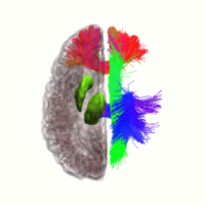
The app contains neuroimaging data from a 33 year old healthy human male brain including:
- 84 cortical areas based on gray matter segmentation of anatomical T1-weighted MRI image (T1)
- 24 subcortical areas, including ventricles and cerebellum segmented from anatomical T1
- 33 white matter pathways reconstructed using tractography from diffusion weighted MRI image (DWI)
- venous segmentation based on susceptibility weighted MRI image (SWI)
- arterial segmentation based on time of flight MRI image (TOF)
- functional connectivity based on blood oxygen level dependent (BOLD) functional MRI images
Cortical and subcortical regions of interest (ROIs) were segmented automatically using the Freesurfer software package:
https://www.sciencedirect.com/science/article/pii/S1053811912000389
White matter pathways were processed and automatically segmented using in-house software developed at Imeka, based on the following publication:
https://www.sciencedirect.com/science/article/pii/S1053811917305839
Venous and arterial segmentations were segmented automatically using in-house software developed at the University of Sherbrooke, based on the following publication:
https://onlinelibrary.wiley.com/doi/abs/10.1002/hbm.24337
All data was acquired with informed consent on a 3 Tesla Philips Ingenia MRI scanner at the Centre Hospitalaire Universite de Sherbrooke in Sherbrooke, Quebec, Canada.
Useful for learning about the anatomy of the brain.